Faculty of mechanic
University of Maribor
Rules
Only words with 2 or more characters are accepted
Max 200 chars total
Space is used to split words, "" can be used to search for a whole string (not indexed search then)
AND, OR and NOT are prefix words, overruling the default operator
+/|/- equals AND, OR and NOT as operators.
All search words are converted to lowercase.
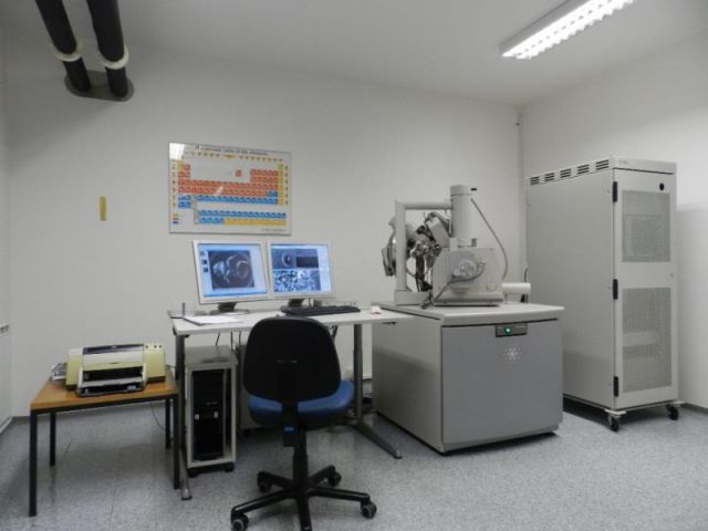
FEI Quanta 200 3D
Environmental scanning electron microscope with tungsten cathode as an electron source (electron beam generation by heat emission).
The microscope is called "environmental" or "ESEM" because it allows work at different pressures and at 100% humidity. By adjusting the pressure in the chamber, we obtain the conditions for observing different samples.
Basic technical characteristics:
- the source of electrons is a tungsten (W) electrode, which allows for a resolution of:
- 3.5 nm at 30 kV (high vacuum)
- 3.5 nm at 30 kV (ESEM mode) <15 nm at 3 kV (low vacuum);
- accelerating voltage from 200 V to 30 kV;
- magnifications from 10 X to 300 000 X;
- five motorized axes (x = 50 mm, y = 50 mm, z = 25 mm, tilt (-10 to 60 degrees) and rotation (n x 360 degrees));
- the size of the chamber equipped with an infrared camera is 379 mm from left to right;
- the source of the ion beam is gallium liquid metal;
- the resolution of the ion gun is 10 nm at 30 kV.
Technical information
With the Quanta 200 3D electron microscope, three basic modes of operation are possible:
- High vacuum - allows the observation of conductive samples (eg metal samples or non-conductive samples coated with a conductive layer (Ag, C or Au)).
- Low vacuum - allows the observation of conductive as well as non-conductive samples without prior preparation (polymeric materials, ceramics, non-conductive surface layers, some biological and medical samples,...). A low vacuum prevents the sample from being electrically overcharged. The pressure can be continuously varied from 1 to 130 Pa.
- In »ESEM« mode, a gas is added to the chamber at low vacuum, which has a low ionization energy and allows the formation of additional "environmental" secondary electrons. These electrons improve the resolution, which is worse at low vacuum than at high vacuum. The gas usually added to the chamber is water vapor.
In the »ESEM« mode we can observe all types of samples (polymeric materials, ceramics, non-conductive surface layers, biological and medical samples,…), but it is especially important for the observation of wet, greasy and dirty samples, and for the observation of in-situ processes (hydration, dissolution…).
Due to different operating conditions, there are several different detectors in the microscope:
- the classic Everthart-Thornley photomultiplier (SEI) for observing morphology and other surface characteristics, capturing secondary electrons in a high vacuum;
- with the backscattered electron detector (BSE) we obtain the contrast between the regions of the sample with different chemical composition (Z-contrast).
- the LF-GSED (Large Field Gaseous Secondary Electron Detector) is suitable for low vacuum operation and the GSED (Gaseous SED) mode is suitable for ESEM.
- with additional equipment built into the microscope (Peltier holder, thermocouple incandescent holder or freezer unit) it is possible to directly observe and photograph in-situ processes. Direct observation is made possible by a special vacuum system that effectively removes products generated during the process and by special detectors for "environmental" secondary electrons.
Ion beam gun
The standard equipment of the Quanta 200 3D microscope is an ion beam gun - FIB (Focused Ion Beam), with which we obtain a focused beam of primary gallium ions. The diameter of the ion beam is very small - (eg 6 nm at a current of 1 pA) and due to the high density of ions allows for a good contrast image.
The basic tasks of an ion beam are:
- etching the surface of the sample;
- cutting;
- polishing the cut surface;
- etching various patterns;
- cutting samples for TEM;
It also provides a high-resolution electron image (by capturing secondary electrons caused by the ion beam). With the ion beam we can process all types of samples (very hard - diamond, metals, ceramics, as well as polymers, medical and biological samples).
The etching rate depends on the sample and the ion flux (Quanta 200 3D has 12 levels - from 1pA to 20 nA).
Electron microscopes have only recently been equipped with ion guns. The applicability of the ion gun in various fields is still the subject of intensive research. It definitely allows us to look below the surface of the sample. The surface is moderately contaminated with Ga ions, which must be taken into account in the microchemical analysis.
Platinum deposition system
Non-standard equipment on the Quanta 200 3D microscope is a platinum deposition system. Platinum deposition has different purposes:
- protect the surface from damage that would occur during ion cutting;
- allows for a more accurate cut;
- reduce or eliminate the electrical charge of the non-conductive sample;
- enable conductive connections between elements (semiconductor industry);
- enables the production of 3D shapes of micron sizes;